How Wireless Handheld Fundus Camera for Eye Exams Works
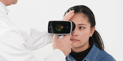
The Wireless handheld fundus camera is an innovative device designed for the examination of the retina, optic nerve head, and other interior structures of the eye. It's an advanced instrument capable of capturing high-resolution images of the retina with extreme accuracy and speed. In this article, we'll explore how the Wireless handheld fundus camera works, its key components, and working mechanisms.
Key Components
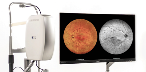
The Wireless handheld fundus camera is made up of several critical components, including the lens, imaging sensor, display screen, and wireless connectivity. The camera's lens is an essential component that allows light to enter the eye, enabling it to take clear and crisp images of the retina. The imaging sensor records these images, converts them into digital signals, and transmits them to the display screen for interpretation. The wireless connectivity enables the operator of the device to control it remotely and send the captured images to a computer or cloud-based storage via Wi-Fi or Bluetooth.
Working Mechanism
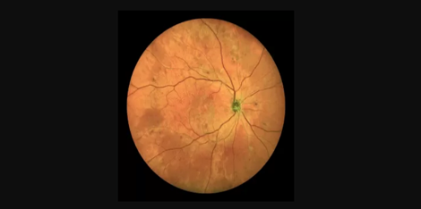
The Wireless handheld fundus camera works by projecting a beam of light into the eye and capturing its reflection from the retina. The device's lens and imaging sensor work together to focus on the retina's interior structures and capture high-resolution images that reveal the optic nerve head, macula, and other retinal landmarks. The camera's display screen allows the operator to view the images clearly in real-time, and the wireless connectivity allows for remote operation and transmission of the images to devices connected via the cloud.
Advanced Technology
The Wireless handheld fundus camera is an essential tool in modern manufacturing due to its advanced technology and efficient production processes. The device's high-resolution imaging capabilities, wireless connectivity, and remote operation features make it an invaluable tool for healthcare professionals, manufacturing companies, and researchers. It enables them to take quick and accurate images of the retina, carry out detailed analyses, and share the results with other stakeholders remotely.
Application Scenarios
The Wireless handheld fundus camera finds practical applications in various settings, including ophthalmic clinics, hospitals, research labs, and manufacturing facilities. It's an essential tool for primary eye care and diagnosis of ocular diseases such as diabetic retinopathy, age-related macular degeneration, and glaucoma. Manufacturing companies use it in quality control processes to ensure the reliability and safety of their products, while researchers use it to study the development and progression of ophthalmological disorders.
Conclusion
The Wireless handheld fundus camera is a crucial device in modern ophthalmology, manufacturing, and research. Its advanced technology, high-resolution imaging, and wireless connectivity features make it an invaluable tool for healthcare professionals, manufacturers, and researchers worldwide. For more information about Wireless handheld fundus cameras and suppliers, please feel free to contact us.

 vision screener