How Ultra Widefield Fundus Camera with ICGA Works
The ultra widefield Fundus Camera with ICGA (Indocyanine Green Angiography) is an advanced technology used for diagnostic imaging of the retina. It is a powerful tool used to diagnose and monitor various eye diseases such as age-related macular degeneration, diabetic retinopathy, glaucoma, and retinal detachment.
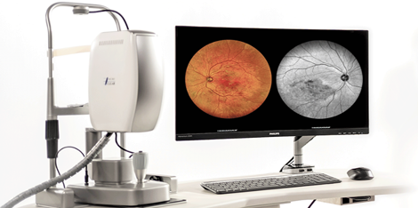
Key Components and Working Mechanisms
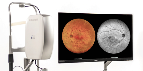
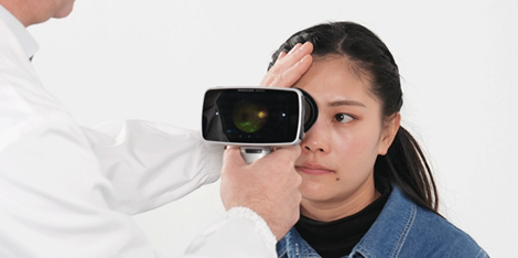
The ultra widefield Fundus Camera with ICGA works by emitting infrared and white light that penetrate through the eye's cornea and lens, reaching the retina. The light reflected from the retina is captured by the camera, generating a high-resolution image of the retina.
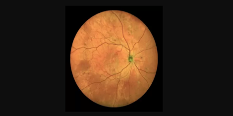
One of the key components of the ultra widefield Fundus Camera with ICGA is the specialized lens system that provides a large view of the retina, allowing for the imaging of peripheral areas of the retina that were previously inaccessible. This significantly improves diagnostic accuracy, especially in conditions such as diabetic retinopathy, where peripheral damage is often the first sign of the disease.
Furthermore, the ultra widefield Fundus Camera with ICGA integrates advanced ICGA technology. ICGA is a diagnostic technique that uses a specialized dye to visualize the blood vessels in the retina. The dye is injected into the patient's arm, and as it circulates throughout the body, it is taken up by the blood vessels in the eye, making them visible to the camera.
Application of Advanced Technology
The ultra widefield Fundus Camera with ICGA is a prime example of how advanced technology can enable efficient production processes. Its specialization in imaging a large view of the retina leads to an accurate and efficient diagnosis of several eye diseases.
Practical Examples or Application Scenarios
A practical application scenario for the ultra widefield Fundus Camera with ICGA could be seen in the diagnosis of age-related macular degeneration. Early symptoms of this condition are difficult to detect as they occur in the peripheral areas of the retina, where normal fundus cameras cannot capture. With the ultra widefield Fundus Camera with ICGA, physicians can detect early symptoms and initiate management before it becomes chronic.
Contact Us and Suppliers
In conclusion, the ultra widefield Fundus Camera with ICGA is an excellent tool for imaging of the retina in eye diseases diagnosis and management. If you are in need of this equipment, contact us to provide you with the necessary information. Additionally, you could also reach out to our suppliers for an effective and efficient delivery.

 Ultra-widefield retinal camera for eye disease dia