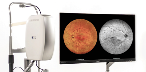
A Retinal Screening Device is a medical device used to assess the health of the retina and detect various eye conditions, including retinal diseases, diabetic retinopathy, macular degeneration, and glaucoma. These devices are essential tools for early detection of eye conditions that could lead to vision loss, enabling timely intervention and treatment.
Retinal screening devices use various imaging techniques to capture detailed images of the retina, allowing healthcare professionals to monitor eye health and diagnose any abnormalities.
Key Types of Retinal Screening Devices:
-
Fundus Cameras:
- Fundus Photography: These devices capture high-resolution images of the retina, including the optic disc, macula, and blood vessels. The images help detect and monitor conditions such as diabetic retinopathy, glaucoma, and age-related macular degeneration.
- Types:
- Color Fundus Camera: Captures color images of the retina to observe the health of blood vessels, the macula, and the optic nerve.
- Red-Free Fundus Camera: Specializes in capturing images with reduced red light, highlighting vascular and nerve fiber layer changes, useful for detecting early diabetic retinopathy and glaucoma.
- Advantages: Non-invasive, quick, and relatively easy to use. Provides clear, detailed images of the retina.
-
OCT (Optical Coherence Tomography):
- Imaging Technique: OCT is a non-invasive imaging technique that uses light waves to take cross-sectional images of the retina. It provides high-resolution images of the retina’s layers and is instrumental in diagnosing retinal diseases like macular edema, glaucoma, and retinal vein occlusions.
- Uses: OCT is commonly used for:
- Early detection of macular degeneration.
- Assessing diabetic retinopathy.
- Monitoring glaucoma progression.
- Advantages: High-resolution, detailed images of retinal layers that can detect changes in the retina even before they are visible through traditional fundus photography.
-
Retinal Scanning Devices:
- Scanning Laser Ophthalmoscope (SLO): This device uses lasers to scan the retina and produce detailed images of the retina's surface. It's used for diagnosing conditions like diabetic retinopathy and macular degeneration.
- Advantages: High sensitivity in detecting retinal abnormalities; can capture 3D images for more detailed assessments.
-
Fundus Fluorescein Angiography:
- Fluorescein Dye Injection: This technique involves injecting a fluorescent dye into a patient’s vein. As the dye circulates through the blood vessels of the retina, a camera captures images of how the dye flows through the retinal blood vessels, helping to identify blockages, leaks, or other retinal issues.
- Uses: Primarily used for detecting conditions like diabetic retinopathy, retinal vein occlusion, and macular degeneration.
- Advantages: Provides a dynamic view of retinal blood vessels and their function.
-
Non-Mydriatic Fundus Cameras:
- No Pupil Dilation: Unlike traditional fundus cameras, non-mydriatic fundus cameras do not require pupil dilation, making them more convenient for patients, particularly those with sensitivities to dilation.
- Advantages: Ideal for screening programs and general check-ups, as it is faster and less uncomfortable for patients.
-
Portable Retinal Screening Devices:
- Portable Fundus Cameras: Smaller, lighter, and more compact, these devices can be used for screening in remote or underserved areas where access to traditional, larger screening equipment may be limited. They provide high-quality images for diagnosis and can be connected to laptops or mobile devices for remote consultations.
- Advantages: Mobility, ease of use, and the ability to perform screening in various settings, including clinics, homes, and field visits.
-
Retinal Tomography (RT):
- Three-Dimensional Imaging: This technology uses light and computer algorithms to create 3D images of the retina and macula. It is often used for diagnosing conditions like macular degeneration, diabetic macular edema, and other retinal pathologies.
- Advantages: Provides detailed 3D imaging that allows for precise diagnosis and monitoring of retinal conditions.
Would you like further information on a specific type of retinal screening device, or are you interested in particular features, models, or brands?
Benefits of Retinal Screening Devices:
Early Detection of Diseases: Retinal screening devices enable early detection of conditions like diabetic retinopathy, macular degeneration, glaucoma, and retinal detachment, which can prevent or minimize vision loss when treated early.
Non-Invasive: Most retinal screening methods are non-invasive and painless, making them more comfortable for patients compared to other medical procedures.
Convenience and Speed: Many retinal screening devices, especially non-mydriatic and portable devices, offer quick and easy screening processes that don’t require significant preparation.
Monitoring and Tracking Progression: Regular screening using these devices allows clinicians to monitor the progression of conditions, adjust treatment plans, and track the effectiveness of treatments over time.
Telemedicine Integration: Some modern retinal screening devices can integrate with telemedicine systems, allowing eye care professionals to analyze images remotely and provide consultations even in remote areas.
Common Retinal Diseases Detected by Retinal Screening Devices:
Diabetic Retinopathy: A complication of diabetes that affects the blood vessels in the retina. Early stages may show no symptoms, but with advanced imaging, changes such as hemorrhages, swelling, or leakage can be detected.
Macular Degeneration: A condition that affects the central part of the retina (macula), leading to loss of central vision. OCT is particularly useful for early detection and monitoring.
Glaucoma: A condition where increased intraocular pressure can damage the optic nerve, leading to peripheral vision loss. Retinal imaging can help detect early changes in the optic nerve and retinal nerve fiber layer.
Retinal Detachment: A medical emergency where the retina separates from its underlying tissue. Early detection can prevent permanent vision loss.
Retinal Vein Occlusion: A blockage of the retinal veins that can lead to vision loss. Fluorescein angiography is commonly used to detect this condition.
Macular Edema: Swelling of the macula, which can result from various conditions like diabetic retinopathy. OCT is effective in detecting macular edema and monitoring its treatment.
Hypertensive Retinopathy: Changes in the retina caused by high blood pressure, which can be detected through fundus photography and angiography.


