How High-Resolution Fundus Camera with ICGA Works
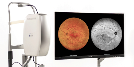
A fundus camera is an important tool in ophthalmology, used to image the retina and optic nerve head. The High-resolution Fundus Camera with Indocyanine Green Angiography (ICGA) is an advanced tool that combines high-resolution imaging with the use of a contrast agent to image blood vessels in the choroid.
Key Components
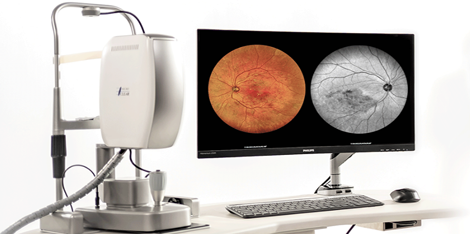
The High-resolution Fundus Camera with ICGA consists of a camera system, a light source, and a computer. The camera system comprises a camera head with a charge-coupled device (CCD) sensor, a lens system, and a mirror. The light source comprises an illumination system that provides white light for imaging the retina and a green laser for ICGA.
Working Mechanisms
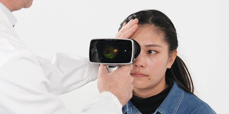
To use the High-resolution Fundus Camera with ICGA, the patient first receives an intravenous injection of indocyanine green, which is a fluorescent dye that emits light when excited by the green laser. The camera head is then positioned close to the patient’s eye, and the mirror is used to focus the light onto the retina. The camera captures the reflected light, which is then sent to the computer for processing.
The computer processes the images obtained by the camera and generates a high-resolution image of the retina. The ICGA images are obtained by illuminating the choroidal vessels with the green laser, causing the ICG to fluoresce. The camera captures the fluorescence emitted by the ICG, enabling the visualization of the choroidal vasculature.
Application of Advanced Technology
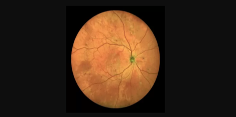
The High-resolution Fundus Camera with ICGA is an example of how advanced technology can improve the practice of medicine. The use of contrast agents and high-resolution imaging allows for the early detection of eye diseases, such as age-related macular degeneration, diabetic retinopathy, and choroidal neovascularization.
Importance in Modern Manufacturing
The importance of the High-resolution Fundus Camera with ICGA in modern manufacturing cannot be overstated. Efficient production processes enable the manufacturer to produce high-quality devices in large quantities, which helps to reduce the cost of the device. This cost reduction, in turn, makes the device more accessible to patients and healthcare providers worldwide.
Practical Examples/Applications
The High-resolution Fundus Camera with ICGA is widely used in ophthalmology clinics and hospitals worldwide. It is an essential tool for the diagnosis, monitoring, and treatment of retinal and choroidal diseases. The device is also used in research to study the pathophysiology and treatment of eye diseases.
Contact Us/Suppliers
If you are interested in purchasing the High-resolution Fundus Camera with ICGA, please contact us. We have a team of experts who can answer any questions you may have about the device. Our suppliers are located worldwide, ensuring that you can purchase the device at a location near you.

 Ultra-widefield retinal imaging camera for diabeti