Fundus Retinal Camera with ICGA for Choroidal Imag: How it Works
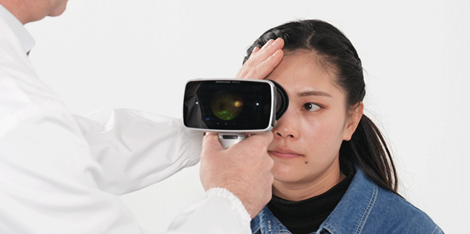
The Fundus Retinal Camera with ICGA for Choroidal Imag is a state-of-the-art technology used in ophthalmology to capture detailed images of the eye's retina and choroid. This instrument is equipped with a specialized camera and a fluorescein angiography system designed to produce images of the choroidal blood vessels.
Key Components and Working Mechanism
The Fundus Retinal Camera with ICGA for Choroidal Imag comprises of an advanced digital camera, digital imaging software, and a fluorescein angiography system. The camera is a powerful imaging tool that enables the technician to capture high-resolution images of the retina and choroid easily.
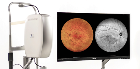
The digital imaging software works with the captured images to produce three-dimensional maps of the choroidal vasculature, helping to diagnose specific eye diseases. Additionally, the fluorescein angiography system employs a unique imaging technique to assess the blood vessels' leakage and blockages. The instrument injects a fluorescent dye inside the patient's bloodstream, before taking a series of images at different intervals.
In each interval, the camera captures the dye's flow in the choroidal blood vessels and provides a detailed view of their architecture. The system also analyzes the images for specific features and structures, enabling the eye care professional to monitor progression or regression of the eye disease accurately.
Application of Advanced Technology
The Fundus Retinal Camera with ICGA for Choroidal Imag's advanced technology enables efficient production processes and accurate medical diagnosis. The technology is easy to use and provides reliable and objective information about the patient's ocular health. This technology has revolutionized and improved the diagnosis and treatment of several eye diseases, including age-related macular degeneration and diabetic retinopathy.
Practical Examples or Application Scenarios
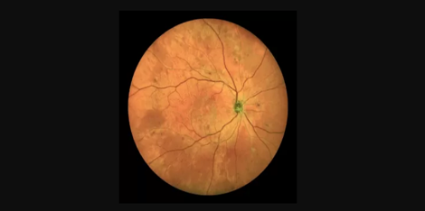
A practical application of the Fundus Retinal Camera with ICGA for Choroidal Imag is in the diagnosis of patients with age-related macular degeneration. By using the fluorescein angiography system, eye care professionals can observe the leakage and blockages in the choroidal vasculature, which are among the leading causes of vision loss in older adults. This information enables the physicians to develop an appropriate treatment plan that can prevent further vision loss.
Contact Us
To learn more about the Fundus Retinal Camera with ICGA for Choroidal Imag and its applications, do not hesitate to contact us. We offer reliable and efficient services that are designed to meet our clients' unique needs.
Suppliers
As a supplier of the Fundus Retinal Camera with ICGA for Choroidal Imag, we provide quality instruments at an affordable price. Contact us today to learn how we can help improve your eye care practice.

 Handheld ultra-widefield color retinal camera