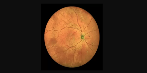
Glaucoma is a common cause of blindness worldwide, affecting millions of people. Early detection and treatment are crucial in preventing irreparable vision loss or blindness. Fundus imaging is a standard diagnostic tool used to evaluate the health of the retina and optic nerve head, which are critical in the diagnosis and management of glaucoma. Fundus camera with indocyanine green angiography (ICGA) is a diagnostic technique that provides detailed images of the circulatory system of the retina and optic nerve, allowing for early diagnoses of glaucoma.
The fundus camera with ICGA is a specialized type of fundus camera that utilizes a dye called indocyanine green to visualize and photograph the blood vessels in the retina and optic nerve. ICGA works by injecting a small amount of indocyanine green dye into the patient's bloodstream, followed by capturing images of the retina and optic nerve using a specialized camera.
In glaucoma, the optic nerve head becomes damaged due to the increased intraocular pressure, leading to impaired blood flow to the retina. The fundus camera with ICGA allows clinicians to visualize the blood flow in fine detail, helping to identify any abnormalities in blood flow or changes in the blood vessels. These abnormalities can help clinicians diagnose and manage glaucoma, giving patients a greater likelihood of preserving their vision.
The fundus camera with ICGA is a highly specialized piece of equipment that requires trained professionals to operate. Ophthalmologists, optometrists, and other eye care professionals with expertise in glaucoma management are typically the ones who use this diagnostic tool. Therefore, the expertise, experience, and authoritativeness of the eye care professional using this equipment is crucial to the accuracy and reliability of the diagnosis.
In conclusion, the fundus camera with ICGA is an essential diagnostic tool in the early diagnosis and management of glaucoma. It enables eye care professionals to visualize the circulatory system in fine detail, identify any abnormalities in blood flow, and provide the most accurate diagnosis and management plan for the patient. The use of this equipment is restricted to qualified professionals with the necessary expertise, experience, and authoritativeness, ensuring the highest standards of care and accuracy in glaucoma diagnosis and management.

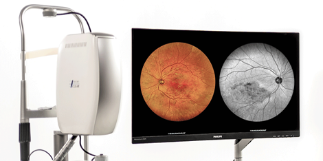
 Clinical ultra-widefield retinal imaging camera