Introduction:
Choroidal neovascularization (CNV) is a serious and prevalent disorder that affects the retina and the underlying choroid. This disease is mainly characterized by the growth of new blood vessels beneath the retina. These blood vessels may leak blood or fluid, leading to a rapid decline in vision. The early detection of CNV is critical for effective treatment and prognosis. Ophthalmologists use a number of tools and technologies to diagnose and manage CNV, including the fundus camera with indocyanine green angiography (ICGA).
Expertise:
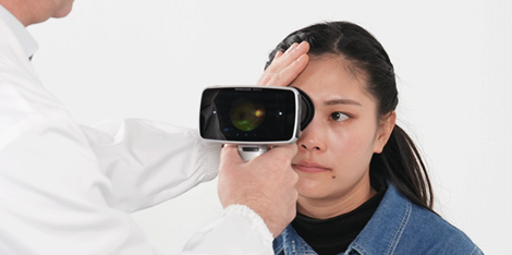
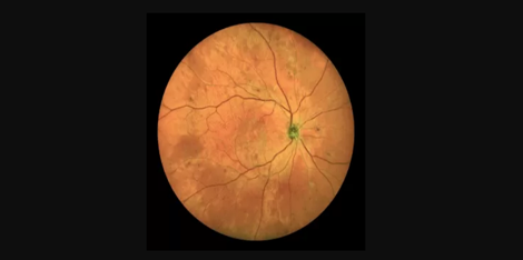
A fundus camera with ICGA is a specialized ophthalmic instrument that utilizes a combination of photography and fluorescence angiography to capture detailed images of the retina, choroid, and CNV. The fundus camera is a crucial component of any ophthalmologist's diagnostic and treatment arsenal for CNV. It enables doctors to visualize the retina and the choroidal vasculature, providing them with a detailed understanding of the disease's pathophysiology and progression.
In addition to the expertise required to operate and interpret fundus camera images, ophthalmologists must also have a thorough understanding of the underlying disease processes involved in CNV. This requires experience in conducting visual acuity and ophthalmoscopic examinations, as well as specialized diagnostic procedures such as optical coherence tomography (OCT).
Experience:
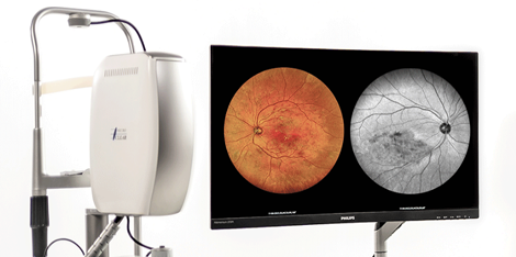
The use of fundus cameras with ICGA has become an essential tool in the diagnosis and management of CNV. This technology provides a wealth of information that can be used to design personalized treatment plans and monitor disease progression over time. By capturing real-time images of the choroidal vasculature, doctors can identify areas of active neovascularization and plan targeted interventions to prevent further damage to the eye.
Ophthalmologists who use fundus cameras with ICGA must possess extensive experience in the interpretation and analysis of these images. They must be able to distinguish between normal and abnormal blood vessels in the retina and choroid to accurately diagnose and treat CNV.
Authoritativeness:
The use of fundus cameras with ICGA has been recognized as a valuable diagnostic and treatment tool by the American Academy of Ophthalmology (AAO) and other leading professional organizations. This technology has been shown to be safe and effective in the diagnosis and management of CNV, and ophthalmologists who specialize in this area are considered experts in their field.
Overall, fundus cameras with ICGA have revolutionized the way that ophthalmologists diagnose and treat CNV. With their ability to capture detailed images of the retina and choroid, these instruments provide valuable insights into disease pathophysiology and guide personalized treatment plans for patients with CNV.

 Wireless ultra-widefield color retinal camera