How Digital Slit Lamp with Image Capture and Storage Works
Digital slit lamp with image capture and storage is an innovative technology that enables the capture and storage of digital images of the human eye. This device is an advanced version of the traditional slit lamp microscope and is widely used in ophthalmology for the examination of the anterior and posterior segments of the eye. In this article, we will delve into how digital slit lamp with image capture and storage works, explaining its key components and working mechanisms.
Working Mechanisms
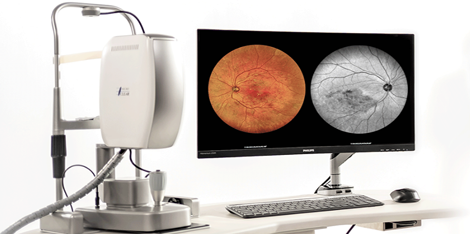
A digital slit lamp is made up of the following components: the microscope, the slit lamp, the camera, and the software. The microscope enables the magnified observation of the eye structure, while the slit lamp provides illumination of the eye. The camera captures high-resolution digital images of the eye structure, which are then stored in a computer's hard drive or server. The software enables the physician to view, edit, and store the captured images.
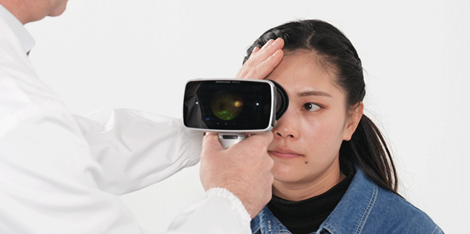
During an eye examination, the physician positions the patient in front of the digital slit lamp, where the anterior and posterior segments of the eye are examined. The physician can view the eye through the microscope and use the slit beam to illuminate the eye structure. The physician can adjust the slit beam's width, height, and angle depending on the eye structure's area that needs to be examined.
The camera, which is attached to the slit lamp, captures images of the illuminated eye. The physician can adjust the camera's focus, aperture, and shutter speed to achieve the desired image quality. The captured images are then stored in the computer's hard drive or server.
Applications of Digital Slit Lamp with Image Capture and Storage
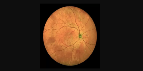
Digital slit lamps with image capture and storage are widely used in ophthalmology for the diagnosis and management of various eye conditions. An example is the detection and grading of diabetic retinopathy, a leading cause of blindness in adults. The captured images of the retina can be graded for evidence of diabetic retinopathy and monitored over time to detect any changes in the eye's condition.
Another application is the detection and management of glaucoma, a progressive eye disease that damages the optic nerve. The captured images of the optic nerve can be used to monitor the progression of the disease and the effectiveness of treatment.
Conclusion
The Digital slit lamp with image capture and storage is an innovative technology that has revolutionized the diagnosis and management of various eye conditions. Its key components, including the microscope, slit lamp, camera, and software, work together to enable the capture and storage of high-resolution digital images of the eye structure. The technology has practical applications in various areas, including diabetic retinopathy and glaucoma management. If you are interested in purchasing this technology, contact us for more information on suppliers and pricing.
Contact us at info@example.com or check out our website for a list of suppliers.


 Ultra-widefield Color Retinal Cameras