Compact Fundus Camera with ICGA: A Revolution in Ophthalmology
The Compact Fundus Camera with ICGA is a technological innovation that has revolutionized the diagnosis and treatment of retinal diseases. It is a non-invasive, reliable and efficient imaging system that produces high-resolution images of the retina in real-time. The system combines color fundus photography with Indocyanine Green Angiography (ICGA) to provide detailed information about the structure and function of the retina. In this article, we delve into how Compact Fundus Camera with ICGA works, explaining its key components and working mechanisms.
Key Components
The Compact Fundus Camera with ICGA is a sophisticated imaging system that comprises several important components. These include:
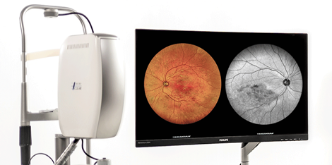
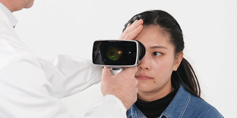
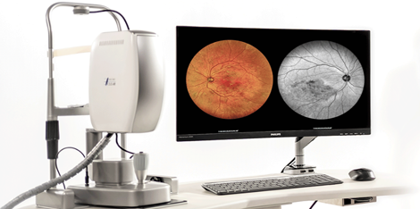
1. Camera Head: The camera head is the most critical part of the Compact Fundus Camera with ICGA. It contains a digital camera sensor that captures high-resolution images of the retina. Its advanced optical design ensures that images are captured with minimal distortion and aberration.
2. Laser Source: The ICGA process requires the use of a laser source to excite the Indocyanine Green dye, which fluoresces at a specific wavelength. The laser source is precisely calibrated to ensure optimal excitation of the dye.
3. Filters: The camera head has a set of filters that enable the capturing of images in different light spectra, such as white light for color fundus photography and infrared light for ICGA.
Working Mechanisms
The Compact Fundus Camera with ICGA works by a combination of different mechanisms. These include:
1. Image Capture: The camera head is mounted on a support structure that is precisely positioned to capture high-resolution images of the retina. Images are captured in real-time and displayed on a monitor for immediate analysis.
2. Indocyanine Green Angiography: This mechanism involves the injection of Indocyanine Green dye into the patient's blood vessels, which fluoresces when excited by a laser. The imaging system captures the fluorescence images of the retinal blood vessels, enabling a detailed analysis of the retinal circulation.
Applications
The Compact Fundus Camera with ICGA is a versatile imaging system with numerous applications in ophthalmology. These include the diagnosis and treatment of conditions such as macular degeneration, diabetic retinopathy, and retinal vein occlusions, among others.
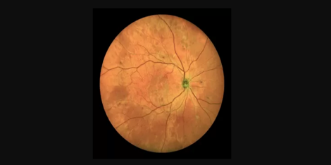
For instance, the Compact Fundus Camera with ICGA is used to detect macular degeneration, which causes the progressive loss of central vision. The system helps to diagnose the disease early and monitor its progression over time. Similarly, the Compact Fundus Camera with ICGA is used to diagnose and treat diabetic retinopathy, a condition that affects up to 80% of people who have had diabetes for more than 20 years.
Conclusion
The Compact Fundus Camera with ICGA is a high-tech imaging system that has transformed ophthalmology. Its advanced technology enables efficient production processes and ensures the best quality images, which are critical to the diagnosis and management of retinal diseases. To learn more about this system, contact us today or visit our suppliers’ website.
Keywords: Compact Fundus Camera with ICGA, ophthalmology, retinal diseases, diagnosis, Indocyanine Green Angiography, laser source, filters, image capture. Contact us. Suppliers.

 Ultra-widefield retinal imaging camera for telemed